
3D Ultrasounds: Complete Guide to Advanced Prenatal Imaging Technology
For expectant parents, seeing their baby’s image is a monumental milestone. While traditional 2D ultrasounds confirm health, the advanced world of 3D and 4D imaging offers a deeper, more profound experience. At GoldenView Ultrasound, we know that the benefits of these elective scans extend far beyond a keepsake photo; they influence emotional bonding, family preparation, and parental peace of mind.
This guide explores the true advantages of investing in advanced prenatal imaging, highlighting why a 3D/4D/HDLive session is an investment in your family’s emotional journey.
The Psychological Power of Prenatal Bonding
The most significant benefit of 3D/4D ultrasounds is the immediate and powerful emotional connection they forge between parents and their baby.
1. Reducing Anxiety and Fostering Reality
- The Emotional Gap: Early in pregnancy, the baby is an abstract concept. This can sometimes lead to anxiety or a sense of emotional distance, especially for first-time parents or partners who cannot feel fetal movement.
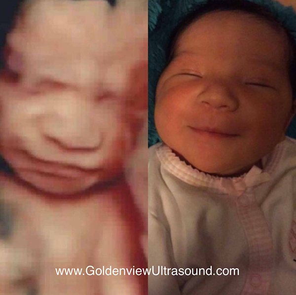
- The Clarity Solution: 3D and HDLive (5D) technology transforms the abstract into the undeniable. Seeing realistic facial features, distinguishing a nose, or catching a tiny yawn makes the baby a distinct person. Studies suggest that this early visual confirmation can reduce prenatal anxiety and make healthy behavioral changes (like improved diet or rest) feel more urgent and important to the mother.
2. Strengthening Paternal and Sibling Bonds
In many medical settings, guests are restricted. This limits the partner’s first visual connection.
- Paternal Connection: For fathers, partners, and other support figures, the 3D/4D scan is often the first moment they truly connect with the baby. Seeing the baby in motion (4D) or recognizing a family trait on the face (3D) accelerates the bonding process, helping them transition into a parenting role sooner.
- Sibling Preparation: Elective sessions allow older siblings to attend and meet their new brother or sister on the big screen. This visual introduction transforms the baby from a distant concept (“Mommy has a baby in her tummy”) into a real, recognizable person, which can significantly reduce sibling rivalry and anxiety before birth.
The Enhanced Detail of Advanced Technology
While GoldenView Ultrasound does not perform diagnostic scans, the superior clarity of our technology offers unique benefits for the parental experience.
1. Visualization of Unique Features
Traditional 2D scans are cross-sections, making it difficult for non-medical professionals to interpret. 3D/4D/HDLive excels at surface rendering:
- Family Traits: The realistic rendering allows parents to clearly identify family resemblances—a fun and heartwarming benefit. Many parents report recognizing a dimple, a nose shape, or a specific movement shared by a parent or sibling.
- Behavioral Glimpses: The real-time motion of the 4D scan captures micro-expressions (frowning, smiling), behavioral patterns (thumb-sucking, stretching), and sleep cycles. These provide a unique “personality preview” that becomes a cherished family memory.
2. High-Value Keepsake Creation
The investment in a 3D/4D session is a long-term investment in memory:
- Video Heirloom: The 4D real-time video is a unique heirloom that can be shared instantly and preserved across generations, capturing motion that a static photo cannot.
- The Heartbeat: The ability to record the baby’s actual heartbeat (for Heartbeat Huggables) transforms the sound into a tangible, comforting object. This unique keepsake is impossible to obtain without the specialized equipment and time dedicated during an elective session.
The Benefit of Cost Transparency and Time
Choosing an elective facility offers practical advantages that reduce stress and maximize comfort, which contributes directly to the quality of the experience.
1. Cost Certainty with Flat-Rate Pricing
In the medical world, surprise bills due to deductibles and facility fees are common.
- Stress Reduction: GoldenView Ultrasound offers flat-rate, transparent pricing. The price quoted is the price paid, eliminating financial stress and allowing parents to budget for this special event with certainty. This straightforward approach enhances the overall relaxation and enjoyment of the session.
2. Time and Comfort
Our sessions are structured around your needs, not medical requirements.
- Dedicated Time: We allocate extended time slots (often 30+ minutes of scan time for premium packages). This is the key to getting great pictures, as it allows the technician to wait patiently for the baby to move into an optimal position, often requiring mom to walk, stretch, or snack.
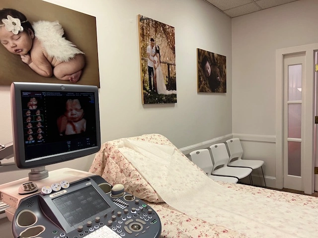
- Family Environment: The spa-like environment, large screens, and open guest policy make the scan an event. The comfort allows the parents and family to fully relax and enjoy the bonding experience.
3. Protection with the “Shy Baby” Guarantee
The ultimate practical benefit is the security of your investment. Because your goal is a clear keepsake image, we guarantee it.
- Free Rescan: If the baby is hiding their face or uncooperative during your 3D/4D session, we offer a repeat visit FREE of charge. This ensures that the only outcome of your investment is a beautiful picture and a happy memory.
The Value of Early Knowledge (DNA and Gender)
For many, the earliest benefit is simply knowing.
1. Early Gender Clarity
- Advanced Timing: Our visual Gender Determination packages start as early as 13 weeks—significantly earlier than many medical offices schedule their routine anatomy scans.
- Fastest Results: For the earliest possible knowledge, our DNA Gender Blood Testing offers 99% accuracy starting at just 6 weeks. This early information is invaluable for planning and sharing the news with family.
2. Family Planning and Preparation
Early and accurate knowledge of the baby’s gender facilitates practical family planning, enabling parents to:
- Finalize nursery colors and themes.
- Purchase or receive necessary items and clothing.
- Host Gender Reveal parties with confidence and accuracy.
Conclusion
The benefits of 3D/4D ultrasounds are deeply personal. They move beyond basic medical necessity to create emotional milestones that last a lifetime. By providing cutting-edge HDLive (5D) technology, prioritizing safety and comfort, and guaranteeing a transparent, flat-rate experience, GoldenView Ultrasound ensures your investment is focused entirely on the joy of meeting your baby.
Ready to see the unseen? Contact us today to schedule your ultimate bonding session.